Gram-negative bacteria are bacteria that do not retain the crystal violet stain used in the gram-staining method of bacterial differentiation. They are characterized by their cell envelopes, which are composed of a thin peptidoglycan cell wall sandwiched between an inner cytoplasmic cell membrane and a bacterial outer membrane.

They are bacteria that define the opposite of gram-positive bacteria in relation to the differential staining technique. During the Gram stain, the gram-negative bacteria will lose the color of the crystal violet dye after an alcohol wash and will take on the pink / red color of the counterstain, safranin.
The two classes of bacteria are differentiated by Gram stain, due to the composition of their cell wall, that is, Gram positive bacteria have a large layer of peptidoglycan and a thin layer of lipid layer, and unlike Gram negative bacteria that have a thick layer of lipids and lack the peptidoglycan layer or some have a very thin layer of the peptidoglycan layer. The absence of peptidoglycan makes its cell wall less strong and therefore the primary stain is easily removed with alcohol and water.
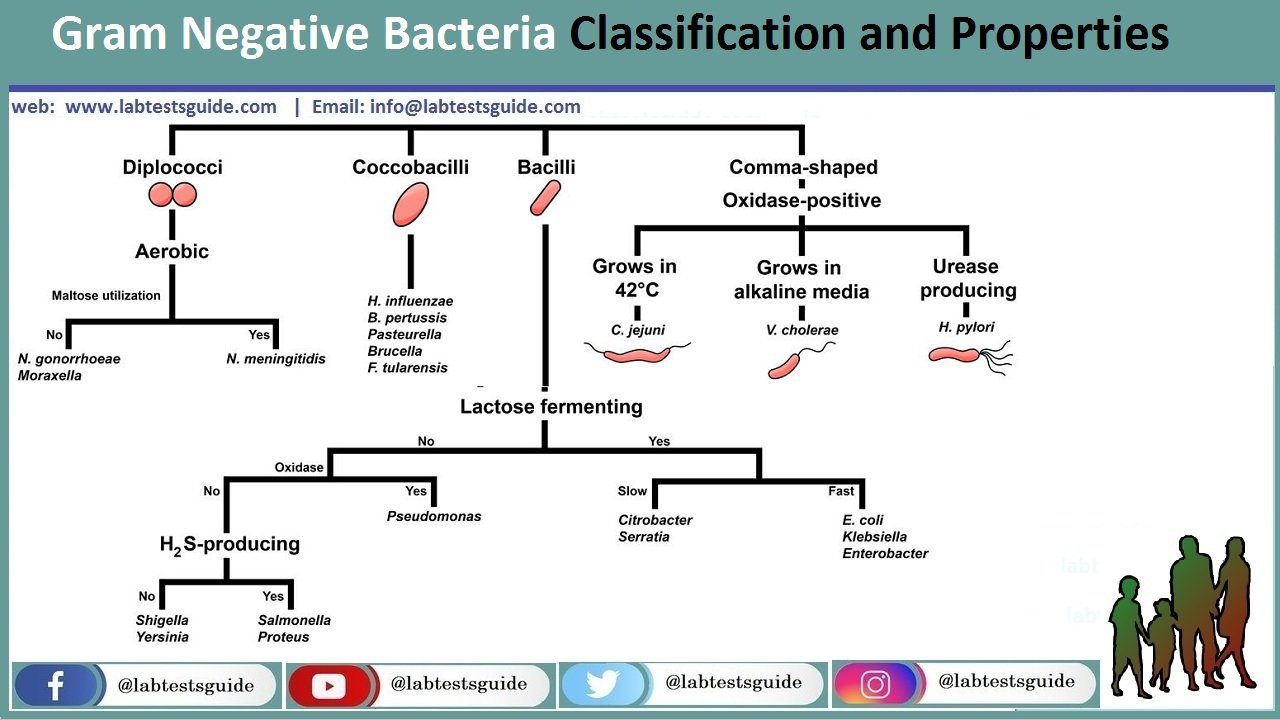
Classifications:
- Diplococci
- Coccobacilli
- Bacilli
- Comma Shaped
1. Diplococci
Aerobic
- N Gonorrhoeae: gonococcus (singular), or gonococci (plural) is a species of Gram-negative diplococci bacteria isolated by Albert Neisser
- Neisseria meningitidis. often referred to as meningococcus, is a Gram-negative bacterium that can cause meningitis and other forms of meningococcal disease such as meningococcemia, a life-threatening sepsis.
2. Coccobacilli
- Haemophilus influenzae: a Gram-negative, coccobacillary, facultatively anaerobic capnophilic pathogenic bacterium of the family Pasteurellaceae.
- Bordetella pertussis: a Gram-negative, aerobic, pathogenic, encapsulated coccobacillus of the genus Bordetella,
- Pasteurella multocida: a Gram-negative, nonmotile, penicillin-sensitive coccobacillus of the family Pasteurellaceae.
- Brucella: a genus of Gram-negative bacteria, named after David Bruce. They are small, nonencapsulated, nonmotile, facultatively intracellular coccobacilli.
- Francisella tularensis: a pathogenic species of Gram-negative coccobacillus, an aerobic bacterium. It is nonspore-forming, nonmotile, and the causative agent of tularemia, the pneumonic form of which is often lethal without treatment.
3. Bacilli
- Lactose fermenting
- Citrobacter: (Slow Lactose fermenting )a genus of Gram-negative coliform bacteria in the family Enterobacteriaceae. The species C. amalonaticus, C. koseri, and C. freundii can use citrate as a sole carbon source. Citrobacter species are differentiated by their ability to convert tryptophan to indole, ferment lactose, and use malonate.
- Serratia: (Slow Lactose fermenting ) Serratia is a genus of Gram-negative, facultatively anaerobic, rod-shaped bacteria of the family Yersiniaceae.
- E. coli : (Fast Lactose fermenting ) Escherichia coli, also known as E. coli, is a Gram-negative, facultative anaerobic, rod-shaped, coliform bacterium of the genus Escherichia that is commonly found in the lower intestine of warm-blooded organisms.
- Klebsiella pneumoniae: (Fast Lactose fermenting ) a Gram-negative, non-motile, encapsulated, lactose-fermenting, facultative anaerobic, rod-shaped bacterium. It appears as a mucoid lactose fermenter on MacConkey agar.
- Enterobacter: (Fast Lactose fermenting ) a genus of common Gram-negative, facultatively anaerobic, rod-shaped, non-spore-forming bacteria of the family Enterobacteriaceae. It is the type genus of the order Enterobacterales.
- Non-lactose fermenting
- Pseudomonas (Oxidase +Ve) is a genus of Gram-negative, Gammaproteobacteria, belonging to the family Pseudomonadaceae and containing 191 validly described species. The members of the genus demonstrate a great deal of metabolic diversity and consequently are able to colonize a wide range of niches.
- Salmonella: (Oxidase -Ve) a genus of rod-shaped Gram-negative bacteria of the family Enterobacteriaceae. The two species of Salmonella are Salmonella enterica and Salmonella bongori. S. enterica is the type species and is further divided into six subspecies that include over 2,600 serotypes.
- Proteus: (Oxidase -Ve) a genus of Gram-negative Proteobacteria. Proteus bacilli are widely distributed in nature as saprophytes, being found in decomposing animal matter, sewage, manure soil, the mammalian intestine, and human and animal feces.
- Shigella: (Oxidase -Ve) a genus of bacteria that is Gram-negative, facultative anaerobic, non-spore-forming, nonmotile, rod-shaped and genetically closely related to E. coli.
- Yersinia: (Oxidase -Ve) a genus of bacteria in the family Yersiniaceae. Yersinia species are Gram-negative, coccobacilli bacteria, a few micrometers long and fractions of a micrometer in diameter, and are facultative anaerobes.
4. Comma Shaped
These are Oxidase Positive:
- C. jejuni: Campylobacter jejuni is one of the most common causes of food poisoning in Europe and in the United States. The vast majority of cases occur as isolated events, not as part of recognized outbreaks.
- V. Cholera: Vibrio cholerae is a Gram-negative, comma-shaped bacterium. The bacterium’s natural habitat is brackish or saltwater where they attach themselves easily to the chitin-containing shells of crabs, shrimps, and other shellfish.
- H. pylori: Helicobacter pylori, previously known as Campylobacter pylori, is a gram-negative, microaerophilic, spiral bacterium usually found in the stomach. Its helical shape is thought to have evolved in order to penetrate the mucoid lining of the stomach and thereby establish infection.
Characteristics:
- Rob/bacillus shaped – Escherichia coli.
- Coccobacillus – which is a combination of both cocci and bacilli shapes include Hemophilus influenza.
- Streptobacillus are rod-shapes that are connected together in chains eg Streptobacillus moniliformis.
- Trichome shape is a series of rod-shaped cells that are arranged in a columnar form and it may be enclosed in a sheath.
- Spiral shaped bacteria also called spirochaetes e.g Chlamydia trachomatis, Treponema pallidum.
- Filamentous shaped gram-negative have a filament-like shape eg Norcadia spp.
Differences Between Gram Positive and Gram Negative Bacteria
Common Differences Between Gram Positive and Gram Negative Bacteria
| Character | Gram-Positive Bacteria | Gram-Negative Bacteria |
|---|---|---|
| Gram Reaction | Retain crystal violet dye and stain blue or purple on Gram’s staining. | Accept safranin afterdecolorization and stain pink or red on Gram’s staining. |
| Cell wall thickness | Thick (20-80 nm) | Thin (8-10 nm) |
| Peptidoglycan Layer | Thick (multilayered) | Thin (single-layered) |
| Rigidity and Elasticity | Rigid and less elastic | Less rigid and more elastic |
| Outer Membrane | Absent | Present |
| Variety of amino acid in cell wall | Few | Several |
| Aromatic and Sulfur-containing amino acid in cell wall | Absent | Present |
| Periplasmic Space | Absent | Present |
| Teichoic Acids | Mostly present | Absent |
| Porins | Absent | Present |
| Lipopolysaccharide (LPS) Content | Virtually None | High |
| Lipid and Lipoprotein Content | Low (acid-fast bacteria have lipids linked to peptidoglycan) | High (because of presence of outer membrane |
| Ratio of RNA:DNA | 8:1 | Almost 1 |
| Mesosomes | Quite Prominent | Less Prominent |
| Flagellar Structure | 2 rings in basal body | 4 rings in basal body |
| Magnetosomes | Usually absent. | Sometimes present. |
| Morphology | Usually cocci or spore forming rods (exception : Lactobacillus and Corynebacterium) | Usually non-spore forming rods (Exception : Neisseria) |
| Endospore formation | Some produce endospores during unfavorable conditions. | Usually not found to produce endospores. |
| Toxin Produced | Exotoxins | Endotoxins or Exotoxins |
| Pathogens | Few pathogenic bacteria belong to Gram positive group. | Most pathogens are Gram negative. |
| Nutritional Requirements | Relatively Complex | Relatively Simple |
| Resistance to Physical Disruption | High | Low |
| Cell Wall Disruption by Lysozyme | High | Low (requires pretreatment to destabilize outer membrane) |
| Susceptibility to Penicillin and Sulfonamide | High | Low |
| Susceptibility to Streptomycin, Chloramphenicol and Tetracycline | Low | High |
| Inhibition by Basic Dyes | High | Low |
| Susceptibility to Anionic Detergents | High | Low |
| Resistance to Sodium Azide | High | Low |
| Resistance to Drying | High | Low |
| Rendering | They can rendered Gram -ve by increasing acidity | They can rendered Gram +ve by increasing alkalinity |
| Examples | Staphylococcus Streptococcus Bacillus Clostridium Enterococcus | Escherichia Salmonella Klebsiella Proteus Helicobacter Pseudomonas |
Related Articles:
RELATED POSTS
View all


