A procedure in which a blood sample is viewed under a microscope to count different circulating blood cells (red blood cells, white blood cells, platelets, etc.) and to see if the cells look normal. Evaluates white blood cells (WBC, leukocytes), red blood cells (RBC, erythrocytes), and platelets (thrombocytes). The blood smear is examined to investigate hematologic problems (blood disorders) and sometimes to look for parasites in the blood, such as malaria and heartworms.
| Also Known as | Peripheral Smear, Blood Film, Red Blood Cell Morphology, Erythrocyte Morphology,Peripheral Blood Smear, Peripheral Blood Picture, RBC Morphology, Blood Smear Analysis,Peripheral Blood Film |
| Test Purpose | This Test that gives information about the number and shape of blood cells |
| Test Preparations | No Need any Preparation |
| Test Components | CBC, Blood Film |
| Specimen | 2 ml EDTA Blood |
| Stability Room | 4 hours |
| Stability Refrigerated | 8 hours |
| Stability Frozen | N/A |
| Method | Microscopy |
| Download Report | Download Report |
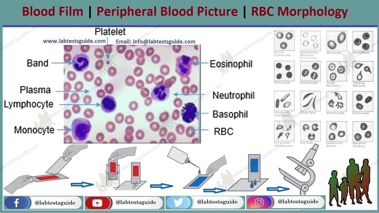
What is a blood smear?
A blood smear is a blood sample that is smeared onto a glass slide that is treated with a special dye. In the past, all blood smears were examined under a microscope by laboratory professionals. Automated digital systems can now be used to help examine blood smears.
The purpose of examining a blood smear is to check the size, shape, and number of three types of blood cells:
Red blood cells, which carry oxygen from the lungs to the rest of the body.
White blood cells, which fight infection.
Platelets, which help blood to clot
Why do I need a blood smear?
You may need a blood smear if you have abnormal results on a complete blood count (CBC). A CBC is a routine test that measures many different parts of your blood.
Your health care provider may order a blood smear if you have symptoms of a blood disorder, such as:
- Fatigue
- Jaundice, a condition that causes the skin and eyes to turn yellow
- unusual bleeding, including nosebleeds
- Fever that lasts, or comes and goes
- Bone-ache
- Anemia
- bruises easily
- A spleen that is larger than normal
Abnormal RBC’s
may be a sign of:
- Anemia
- sickle cell anemia
- Hemolytic anemia, a type of anemia in which the body destroys red blood cells faster than they are replaced
- thalassemia
- Bone marrow disorders
- liver disease
- Cancer that has spread to the bone
Abnormal WBC’s
may be a sign of:
- Infection or Inflammation
- Allergies
- Leukemia
- Bone Marrow Disorders
Abnormal Platelets
may be a sign of:
- Thrombocytopenia, a condition in which the blood does not have enough platelets, increasing the risk of bleeding
- Hereditary platelet disorders (rare), such as Bernard-Soulier syndrome
When to Get Tested :
- A general feeling of tiredness or weakness
- Lack of energy
- Frequent infections
- Fatigue
- Bone and joint pain
- Excessive bleeding and bruising
- Fever
- Weakness
- Weight loss
- Swollen lymph nodes, liver, spleen, kidneys, and testicles
- Frequent infections
- Headaches
- Vomiting
- Confusion
- Seizures (may occur when cells are increased in the brain or central nervous system)
- Night sweat
Sample Required:
2 ml EDTA Blood Sample Required.
Sample Preparations;
No Need any Preparations required
Normal results Mean :
Red blood cells (RBCs) are normally the same size and color and are lighter in color in the center. The blood smear is considered normal if there is:
Normal appearance of cells.
Normal white blood cell differential
Normal value ranges may vary slightly between different laboratories. Some labs use different measurements or test different samples. Talk to your provider about the meaning of your specific test results.
Abnormal Results Mean :
Abnormal results mean that the size, shape, color, or coating of the red blood cells is not normal.
Some abnormalities can be rated on a 4-point scale:
- 1+ means that a quarter of the cells are affected
- 2+ means half of the cells are affected
- 3+ means that three quarters of the cells are affected
- 4+ means all cells are affected
The presence of cells called target cells may be due to:
- Deficiency of an enzyme called lecithin cholesterol acyl transferase
- Abnormal hemoglobin, the protein in red blood cells that carries oxygen (hemoglobinopathies)
- Lack of iron
- liver disease
- removal of the spleen
The presence of sphere-shaped cells may be due to:
- Low number of red blood cells because the body destroys them (immune hemolytic anemia)
- Low number of red blood cells due to some sphere-shaped red blood cells (hereditary spherocytosis)
- Increased breakdown of red blood cells
The presence of oval-shaped red blood cells may be a sign of hereditary elliptocytosis or hereditary ovalocytosis. These are conditions in which the red blood cells are abnormally shaped.
The presence of fragmented cells may be due to:
- Artificial heart valve
- Disorder in which the proteins that control blood clotting become overactive (disseminated intravascular coagulation)
- Infection in the digestive system that produces toxic substances that destroy red blood cells, causing kidney damage (hemolytic uremic syndrome)
- Blood disorder that causes blood clots to form in small blood vessels throughout the body and leads to a low platelet count (thrombotic thrombocytopenic purpura)
The presence of a type of immature red blood cells called normoblasts may be due to:
- Cancer that has spread to the bone marrow
- Blood disorder called erythroblastosis fetalis that affects the fetus or newborn
- Tuberculosis that has spread from the lungs to other parts of the body through the blood (miliary tuberculosis)
- Bone marrow disorder in which the marrow is replaced by fibrous scar tissue (myelofibrosis)
- spleen removal
- Severe breakdown of red blood cells (hemolysis)
- Disorder in which there is excessive breakdown of hemoglobin (thalassemia)
The presence of cells called burr cells may indicate:
- Abnormally high level of nitrogen waste products in the blood (uremia)
The presence of cells called spur cells may indicate:
- Inability to fully absorb dietary fat through the intestines (abetalipoproteinemia)
- severe liver disease
The presence of tear-shaped cells may indicate:
- Myelofibrosis
- Severe iron deficiency
- Thalassemia major
- Bone Marrow Cancer
- Anemia caused by the bone marrow not producing normal blood cells due to toxins or tumor cells (myelophthisic process)
The presence of Howell-Jolly bodies (a type of granule) may indicate:
- The bone marrow does not make enough healthy blood cells (myelodysplasia)
- The spleen has been removed
- sickle cell anemia
The presence of Heinz bodies (pieces of altered hemoglobin) may indicate:
- Alpha thalassemia
- Congenital hemolytic anemia
- Disorder in which red blood cells break down when the body is exposed to certain medications or stressed due to infection (G6PD deficiency)
- Unstable form of hemoglobin
The presence of slightly immature red blood cells may indicate:
- Anemia with bone marrow recovery
- Hemolytic anemia
- Hemorrhage
The presence of basophilic stippling (a mottled appearance) may indicate:
- lead poisoning
- Bone marrow disorder in which the marrow is replaced by fibrous scar tissue (myelofibrosis)
- The presence of sickle cells may indicate sickle cell anemia.
| Terminology | Morphology | Explanation |
| Microcytes | 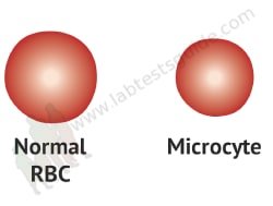 | RBC size is smaller than normal |
| Macrocytes | 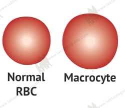 | RBC size is larger than normal |
| Anisocytosis | 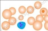 | Variation in RBC size |
| Poikilocytosis | 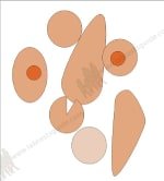 | Variation in RBC shapes |
| Polychromasia | 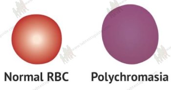 | RBCs are large and basophilic. |
| Spherocytes |  | RBCs are small and round and no central pale area. |
| Elliptocytes | 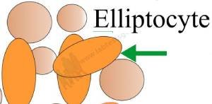 | RBCs are rod-shaped (oval or elliptical) |
| Target cells | 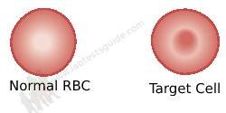 | RBCs have a darkly central area. |
| Tear-drop poikilocyte | 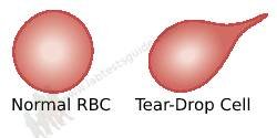 | RBC looks like tears |
| Sickle cells | 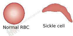 | Moon shaped RBC |
| Acanthocytes | 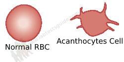 | RBCs have spikes, irregular projections |
| Schistocytes |  | RBCs show fragments and a variety of shapes and sizes. |
| Pencil cells | 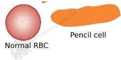 | Elongated RBC |
| Burr cells | 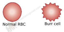 | RBCs are created and irregular. |
| Howell-Jolly bodies |  | RBCs show nuclear remnants. |
| Heinz bodies | 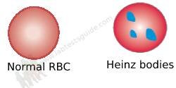 | RBCs show denatured hemoglobin. |
| Toxic granulation | 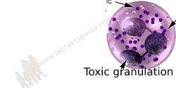 | Increased granules in polys |
| Rouleaux formation | 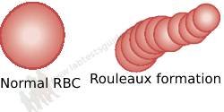 | RBCs show clumping or aggregation. |
| Agglutination of RBCs |  | RBCs are clumped |
Related Content
Keywords: rbc morphology,morphology of rbc,rbc morphology in hindi,rbc morphology reporting,rbc morphology hypochromia,rbc morphology test,rbc morphology meaning in hindi,rbc morphology lab test,morphology,rbc morphology ppt,rbc morphology quiz,rbc morphology normocytic normochromic,rbc morphology abnormal,red blood cell morphology,abnormal red cell morphology,rbc morphology interpretation,abnormal red blood cell morphology,abnormal morphology of red blood cells, peripheral blood smear,blood,peripheral smears,peripheral blood,blood cells,red blood cells,white blood cells,peripheral blood smear microscopy,blood smear,peripheral blood film,peripheral,peripheral blood smear lab,peripheral blood stem cells,peripheral blood smear in hindi,peripheral blood smear practical,peripheral blood smear pathology,how to make peripheral blood smear,peripheral blood smear histology,peripheral blood smear hematology
RELATED POSTS
View all
