A urinalysis is a test of your urine. A urinalysis is used to detect and manage a wide range of disorders, such as urinary tract infections, kidney disease and diabetes.
A urinalysis involves checking the appearance, concentration and content of urine. Abnormal urinalysis results may point to a disease or illness.

Also Known as: Urine Test, Urine Analysis , Urine CE, Urine C/E, UCE, Urinalysis
Test Panel: Physical properties, Chemical Tests, Dipstick Tests, Microscopic Examination
Type of urine samples:
- Random sample:
This is a diluted urine sample and may give an inaccurate interpretation of patient health. But is best to do microscopy to evaluate WBC or RBC. - First Morning sample:
This is the best sample for microscopy and urine analysis. This is the concentrated urine because of urine remained throughout the night in the urinary bladder. This will contains an increased concentration of analytes and cellular elements. Urine must have remained in the bladder for 8 hours is considered as the first-morning sample. - Urine for sugar (Postprandial 2 hours):
Postprandial 2 hours sample collected after 2 hours of high carbohydrate diet. - Midstream clean catch urine:
This sample is needed for the culture and sensitivity of urinary infection. The patient is advised to clean the urethra, then discard the first few mL of urine. Now midstream of the urine is collected in the sterile container. - 24 Hours of a urine sample
- In this case, discard the first urine and note the time.
- Now collect urine in the container for 24 hours and put the last sample in the container.
- Refrigerate the sample.
- This 24 hours samples are needed for measuring urea, creatinine, sodium, potassium, glucose, and catecholamines.
- Suprapubic collection of the urine sample:
This is done in the patients who cannot be catheterized and the sample is needed for culture. This sample is collected by the needle. - Catheter collection of urine:
This is done by patients who are bedridden and can not urinate. - Pediatric urine sample:
In infants, special collection bags are made adherent around the urethra. Then urine is transferred to a container.
Urine Casts:
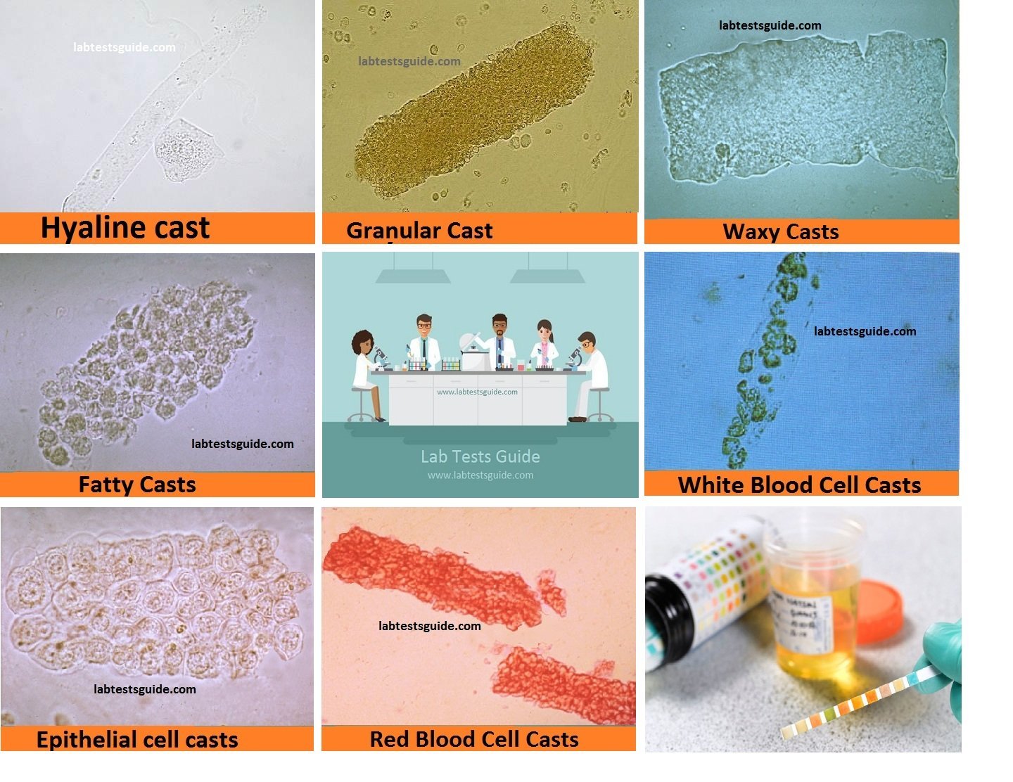
Urinary casts are microscopic cylindrical structures produced by the kidney and present in the urine in certain disease states. They form in the distal convoluted tubule and collecting ducts of nephrons, then dislodge and pass into the urine, where they can be detected by microscopy.
| Acellular casts | Cellular casts |
|---|---|
| Hyaline Casts | Red Blood Cell Casts |
| Granular Casts | White Blood Cell Casts |
| Waxy Casts | Bacterial Casts |
| Fatty Casts | Epithelial Cell Casts |
| Pigment casts | Eosinophilic cast |
| Crystal casts |
- Casts mean inflammation or damage to :
- The tiny tubes in the kidneys.
- Poor blood supply to the kidneys.
- Poisoning (such as lead or mercury).
- Heart failure.
- Bacterial infection.
Summary
| Casts | Composition |
|---|---|
| Hyaline casts | Solidified Tamm-Horsfall mucoprotein |
| Granular casts | Various cell types (Degeneration of cellular casts, Aggregates of plasma proteins or immunoglobulin light chains) |
| Waxy casts (renal failure casts) | Various cell types (Final stage of degeneration of cellular cast) |
| Fatty casts | Lipid droplets within the protein matrix of the cast |
| RBC Casts | Red Blood Cells |
| WBC Casts | White Blood Cells |
| Epithelial Cell Casts | Renal Tubular Epithelial Cells |
| Bacterial Cell Casts | Bacterial Cells |
Hyaline casts:
The most common type of plaster, hyaline plaster, is solidified Tamm-Horsfall mucoprotein secreted by the tubular epithelial cells of individual nephrons. Low urine flow, concentrated urine or an acidic environment can contribute to the formation of hyaline molds and, as such, can be seen in normal people in dehydration or vigorous exercise. Hyaline molds are cylindrical and transparent, with a low index of refraction, so they can be easily lost in the superficial review under bright field microscopy, or in an aged sample where dissolution has occurred, while, on the other hand, Phase contrast microscopy leads to easier identification. Given the ubiquitous presence of the Tamm-Horsfall protein, other types of gypsum are formed by the inclusion or adhesion of other elements to the hyaline base.

Causes
- Normal finding in concentrated urine
Granular casts:
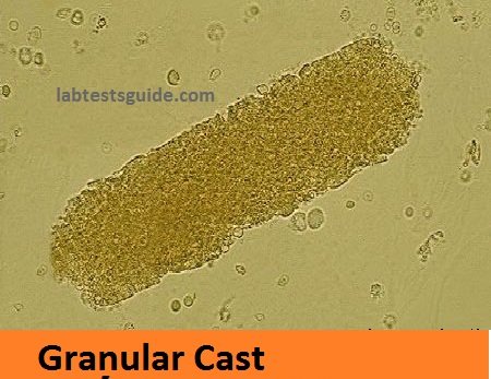
The second most common type of plaster, granular molds may be the result of the breakdown of cell molds or the inclusion of plasma protein aggregates (eg, albumin) or immunoglobulin light chains. Depending on the size of the inclusions, they can be classified as thin or thick, although the distinction has no diagnostic importance. Its appearance is generally more cigar-shaped and with a higher refractive index than hyaline molds. While most of the time it is indicative of chronic kidney disease, these casts, like hyaline casts, can also be seen for a short time after strenuous exercise. The “muddy brown mold” seen in acute tubular necrosis is a type of granular mold.

Causes
- Renal parenchymal Disease
Waxy casts:
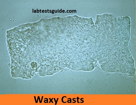
It is believed that they represent the final product of the evolution of the molds, waxy molds suggest the very low flow of urine associated with a serious and long-standing kidney disease, such as kidney failure. In addition, due to urinary stasis and its formation in dilated and diseased ducts, these casts are significantly larger than hyaline casts.
- They are cylindrical.
- They have a higher refractive index.
- They are more rigid and show sharp edges, fractures and broken ends.
Waxy molds are wide molds, which is a more general term to describe the wider molding product of a dilated duct. It is seen in chronic renal failure.
In nephrotic syndrome there are many additional types of cast, including wide and waxy casts if the condition is chronic (this is known as telescopic urine with the presence of many casts).

Causes
- Advanced renal failure
Fatty casts:
Formed by the breakdown of lipid rich epithelial cells, these are hyaline molds with inclusions of yellowish-roasted fatty blood cells. If cholesterol or cholesterol esters are present, they are associated with the “Maltese cross” sign under polarized light. They are pathognomonic for nephrotic syndrome of high urinary proteins.

Causes
- Nephrotic syndrome: Proteinuria
- Primary Lipoid Nephrosis
- Kimmelstiel-Wilson Syndrome
- SLE
- Amyloid
- Subacute Glomerulonephritis
Pigment casts:
Formed by the adhesion of metabolic decomposition products or drug pigments, these molds are named for their discoloration. Pigments include those produced endogenously, such as hemoglobin in hemolytic anemia, myoglobin in rhabdomyolysis and bilirubin in liver disease. Drug pigments, such as phenazopyridine, can also cause discoloration by plaster.
Crystal casts:
Although crystallized urinary solutes, such as oxalates, urates or sulfonamides, can become entangled in a ketanaline mold during their formation, the clinical importance of this fact is not considered to be great.
Red blood cell (RBC) casts:
The presence of red blood cells inside the cast is always pathological and is strongly indicative of granulomatosis with polyangiitis, systemic lupus erythematosus, post-streptococcal glomerulonephritis or Goodpasture’s syndrome. They can also be associated with renal infarction and subacute bacterial endocarditis. They are yellowish brown and are generally cylindrical with sometimes irregular edges; Its fragility makes it necessary to inspect a fresh sample. They are usually associated with nephritic syndromes or urinary tract injuries.

Causes:
- Glomerulonephritis
- Vasculitis
White blood cell (WBC) casts:
Indicative of inflammation or infection, the presence of white blood cells inside or on plasters suggests pyelonephritis, a direct infection of the kidney. They can also be observed in inflammatory conditions, such as acute allergic interstitial nephritis, nephrotic syndrome or acute post-streptococcal glomerulonephritis. White blood cells can sometimes be difficult to distinguish from epithelial cells and may require special staining. Differentiation of simple groups of white blood cells can be done by the presence of hyaline matrix.

Causes
- Interstitial nephritis
- Pyelonephritis
Bacterial casts:
Given their appearance in pyelonephritis, these should be seen in association with loose bacteria, white blood cells and white blood cell cylinders. Its discovery is likely to be rare, due to the efficacy of fighting neutrophil infections and the possibility of misidentification as a fine granular mold.
Epithelial cell casts:
This mold is formed by the inclusion or adhesion of desquamated epithelial cells of the tubule lining. The cells can adhere in random order or in sheets and are distinguished by large, round nuclei and a smaller amount of cytoplasm. These can be seen in acute tubular necrosis and toxic ingestion, such as mercury, diethylene glycol or salicylate. In each case, the groups or sheets of cells can detach simultaneously, depending on the focus of the lesion. Cytomegalovirus and viral hepatitis are organisms that can also cause the death of epithelial cells.

Causes
- Acute Tubular Necrosis (ATN)
- Interstitial nephritis
- Glomrerulonephritis
Eosinophilic cast:
This type of plaster contains eosinophils. It is seen in interstitial nephritis of the tubule and occurs in allergy, commonly to medications such as methicillin and NSAIDs.
Related Articles:
RELATED POSTS
View all
