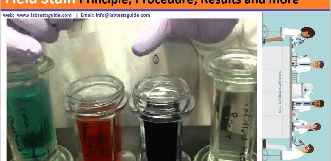
Field stain is a histological method for staining of blood smears. It is used for staining thick blood films in order to discover malarial parasites. Field’s stain is a version of a Romanowsky stain, used for rapid processing of the specimens.

Principle:
- Field’s stains contain methylene blue and eosin.
- These basic and acidic dyes induce several colours when applied to cells.
- The fixator, methanol, does not allow any additional changes to the slide.
- The basic component of peripheral white blood cell( cytoplasm) is stained with acid dye and described as eosinophil or acidophil.
- The acid component that is nucleic acid of the nucleus takes on the basic dye and is stain blue to violet called basophil.
- The neutral components of the cells are stained by both dyes(Field’s stain A and B solution)
Somposition:
Field’s Stain A
| Methylene blue | 1.300 gram |
| Potassium phosphate | 6.250 gram |
| Disodium hydrogen phosphate | 5.000 gram |
| Fresh distilled water | 550.000 ml |
Field’s Stain B
| Eosin | 1.300 gram |
| Disodium hydrogen phosphate | 5.000 gram |
| Potassium dihydrogen phosphate | 6.250 gram |
| Distilled water | 500.000 ml |
- Field’s stain A has is a dark violet coloured solution
- Field’s stain B has is an orange coloured solution
Procedure:
- Place a drop of blood on a slide and spread it over an area of about 1 cm2.( The film should be distributed so thinly that it appears transparent.
- Allow the film to air dry (Do not fix the slide)
- Flood or dip the slide in Field’s Stain A for 2-3 seconds.
- Wash or rinse the slide with distilled water (Agitate gently)
- Flood or dip the slide in Field’s Stain B for 2-3 seconds and wash with distilled water.
- Now air dry the smear or slide and observe under microscope.
Results:
| Neutrophil granules | lilac |
| Eosinophilic granules | orange |
| Red cells | pink |
| Nuclei | blue |
| Malaria parasites | deep red chromatin and pale blue cytoplasm. |
| Leucocyte | purple nuclei and pale blue background |
| Red cells are lysed | only background stroma remains. |
Related Articles:
RSS Error: https://www.labtestsguide.com/category/chemicals/feed is invalid XML, likely due to invalid characters. XML error: > required at line 359, column 16
RELATED POSTS
View all

