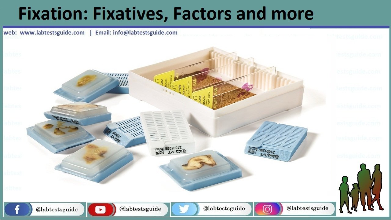
Fixation is a process in which tissue & cells are preserved in a state as close to life as possible. No, micro & macro structures should be lost.
OR
It is the process by which constituent of cells and tissues are fixed in a physical and partly in a chemical state so that they will withstand subsequent treatment with various reagents, with minimum loss, distortion and decomposition.

Fixatives:
Chemical or Physical Agents which are used to prevent and preserve the tissue are called fixatives.
Ideal Fixative:
- It should preserve the tissue volume so that tissue should not change its shape and size.
- To prevent hardness of tissue and should maintains proper tissue consistency.
- It should be non-toxic and non allergic.
- It should penetrate the tissue quickly.
- Tissue should be as near to the living state as possible especially in Research work.
- Should prevent short and long term destruction of tissue by stopping the activity of catabolic enzymes and autolysis and bacterial/fungal attack.
- Nonflammable.
- Should be cost effective.
- Should support good staining with H&E both initially and after storage of the paraffin blocks for at least a decade.
- Should be used for variety of tissues e.g. fatty, lymphoid, and neural tissues.
- It should preserve small and large specimens and support histochemical, immunohistochemical and other specialized procedures.
- They have 1 year of lifespan and also are Stable.
- Compatible with modern automated tissue processors.
- The fixative should be easy to dispose off.
- Support long-term tissue storage giving excellent microtomy of paraffin block.
Factors Involved In Fixation
- Buffers and hydrogen ion concentration
- Temperature
- Penetration
- Osmolality
- Substances added to vehicles
- Concentration of fixative
- Duration of fixation
Buffers and hydrogen ion concentration:
- Normal range for fixative PH 6-8
- Adjusted to the physiological range
e.g. Gastric Mucosa = Acidic 5.5
Esophagus = Neutral 7.0
- For EM glutaraldehyde with increasing pH
- Hypoxia of tissues lowers the pH
- For the prevention of uncontrolled or excessive acidity. There should be buffering capacity into a fixative.
- Some common buffers are bi-carbonate, phosphate, vernol and cacodylate.
- The formalin which is available commercially have pH neutral 7.0 with phosphate buffer.
2. Temperature:
- Surgical specimens are fixed at room temperature.
- For electron microscopy, tissue should be fixed at 0-40’C because at this temperature, the rate of autolysis slow down.
- To fix the blood film and bacteria there should be heat fixation is needed.
- For rapid fixation, formalin is heated at 600’C but the risk of tissue distortion increases.
3. Penetration of fixative:
- Penetration of tissues depends upon the diffusibility of each individual fixative which is a constant.
- It is very important phenomenon of fixation.
- As it is slow process tissue block should be of small size otherwise various zones are formed in the tissue.
- Formalin and alcohol penetration is best.
- Glutraldehyde is worst.
- d=k √ t
- Depth penetrated to the square root of time.
4. Osmolality of the fixative solution:
- Isotonic is normally physiological saline
- Hypertonic solutions cause shrinkage of cells.
- Hypotonic solution cause swelling of cells.
- Slightly hypertonic is ideal.
- For electron microscopy, hypertonic solution is preferable.
5. Concentration of fixative:
- This depends on following factors:
- Cost 2. Availability 3. Effectiveness
- Formalin is best at 10%.
- For EM glutar-aldehyde is generally made up at 0.25% to 4%.
Duration of fixation:
- This depends on the type of fixative e.g. in formaldehyde, duration period is very short otherwise, shrinkage and hardness of tissue will take place.
- In a glutar-aldehyde fixation is prolong.
7. Substances added to vehicles:
- Different substances are added in a fixative to enhance their function e.g. sodium sulphate and NaCl included in a fixative having mercuric chloride.
- NaCl increases the binding of mercuric chloride with protein amino acids.
Secondary fixation:
- Secondary fixation of tissue section is done with iodine soln. to remove black crystals.
- Tissue blocks fixed in gluteraldehyde for EM are commonly fixed osmium tetraoxide.
- Post fixation with dichromate has been recommended for mitochondria.
Fixation Artefacts:
- Formalin pigment is a brown/black pigment formed in tissues fixed with acidic formalin.
- Eliminated by treatment with phenolic formalin.
Materials:
- Fixation:
- Fixative solution (usually commercially available formalin).
- Phosphate buffer (pH = 6.8).
- Rubber or gloves (see Note 1).
- Protective clothing.
- Eyeglasses and mask.
- Fume hood.
- Containers with appropriate lids (volume is commensurate
with sample size. Large neck plastic containers are preferable
and can be reused). - Labels and permanent ink
- Trimming:
- Fume hood.
- Rubber or gloves
- Protective clothing.
- Eyeglasses and mask.
- Dissecting board (plastic boards are preferred as they can be easily cleaned and autoclaved).
- Blunt ended forceps (serrated forceps may damage small animal tissues).
- Scalpels blades and handle.
- Plastic bags and paper towels.
- Containers for histological specimens, cassettes and permanent abels. Containers and cassettes, should be correctly labeled before starting tissue trimming.
- Pre-embedding
- Disposable plastic cassettes for histology (with appropriate
- lids). For small samples, disposable plastic cassettes for histology with subdivision (Microsette®).
- Foam pads (31 × 25 × 3 mm) can be used to immobilize tissue
samples inside the cassettes. - Commercial absolute ethyl alcohol and 96% ethanol solution.
- 90% and 70% ethanol solutions.
- Paraffin solvent/clearing agent: xylene or substitute (e.g.,
Histosol®, Neoclear®). - Paraffin wax for histology, melting point 56–57°C (e.g.,
Paraplast® Tissue Embedding Media). - Automated Tissue Processor (vacuum or carousel type).
- Embedding
- Tissue embedding station (a machine that integrates melted paraffin dispensers, heated and cooled plates).
- Paraffin wax for histology, melting point 56–57°C (e.g. Paraplast® Tissue Embedding Media).
- Histology stainless steel embedding molds. These are available in different sizes (10 × 10 × 5 mm; 15 × 15 × 5 mm; 24 × 24 × 5 mm; 24 × 30 × 5 mm, etc.).
- Small forceps.
- Sectioning:
- Rotary microtome.
- Tissue water bath with a thermometer. Alternatively a thermostatic warm plate can be used.
- Disposable microtome blades (for routine paraffin sections use wedge-shaped blades).
- Sharps container to discard used blades.
- Fine paint brushes to remove paraffin debris.
- Forceps to handle the ribbons of paraffin sections.
- Clean standard 75 × 25 mm microscope glass slides (other dimension microscope glass slides are commercially available).
- Laboratory oven (set at 37°C).
- Coated glass slides (e.g., Superfrost® or Superfrost Plus®). This is especially recommended when slides are used for immunohistochemistry.
- 0.1% gelatin in water (1 g of gelatin in 1 L of distillated water). This should not be used with Superfrost® or Superfrost Plus® slides and should be reserved for immunohistology sections
- Staining and Cover Slipping:
- Harris hematoxylin (commercial solution, ready to use).
- Eosin Y solution.
- Hydrochloric acid 37%.
- Absolute ethanol.
- Ethanol 96%.
- Clearing agent (xylene or substitute e.g. Histosol®, Neoclear®).
- Staining dishes and Coplin jars suitable for staining.
- Permanent mounting medium (e.g. Eukitt®).
- Glass cover slips (25 × 60 mm).
- Filter paper.
- Ethanol solutions:
- Add 12.5 mL of water to 1 L of commercial 96% ethanol to obtain 95% ethanol.
- Add 408 mL of water to 1 L of commercial 96% ethanol to obtain 70% ethanol.
- Storage of Paraffin Blocks and Slides:
- Paraffin blocks storage cabinets.
- Histological slides storage cabinets.
Related Articles:
Keywords: histopathology procedure pdf ,histopathology procedure ppt ,histopathology procedure manual ,histopathology staining procedure ,fish histopathology procedure ,histopathology lab procedures ,histopathology complete procedure ,histopathology slide preparation procedure ,histopathology lab procedure ,histopathology policy and procedure ,procedure for histopathology ,procedure in histopathology ,histopathology laboratory procedure ,procedure of histopathology ,histopathology tissue processing procedure ,routine histopathology procedure ,histopathology staining procedure pdf ,histopathology test procedure
RELATED POSTS
View all

